By: Rochelle McAdam
Date: January 12, 2022
As many of us know from personal experience, cancer is a devastating disease. This is particularly true of a subset of tumours that target the brain and spinal cord regions, collectively known as “gliomas”.
Gliomas are generally considered to fall into two groups, based on how aggressive the tumours are at the time of diagnosis: “low-grade” (associated with longer patient survival time) and “high-grade” (associated with shorter patient survival time). Don’t be fooled by the name “low-grade”, these tumours are capable of transforming into high-grade tumours and can be fatal in both children and adults. The most devastating type of high-grade gliomas are known as glioblastomas, only 5% of patients with these tumours survive five years after their diagnosis and the median survival time is just 12-24 months.
One of the main reasons why gliomas have such poor outcomes is that this disease is very hard to treat. The standard treatment strategy for adult high-grade gliomas involves the surgical removal of the tumour mass, followed by radiation targeted at the brain and chemotherapy. This strategy is used to treat many different types of tumours and is often effective in some cancers, including other types of brain tumours.
The research community is trying to understand why this approach is unsuccessful in glioma patients. Two of the known reasons why this strategy fails are:
1) The successful removal of a brain tumour often depends on which area of the brain the tumour is growing. If it is close to an area important for brain function, it is difficult to operate and completely remove the tumour without damaging that area.
2) Glioma cells can be highly infiltrative. This means that tumour cells can migrate away from the tumour mass and lay dormant in normal brain areas, escaping surgical removal. These cells can then act as a reservoir for tumour regrowth.
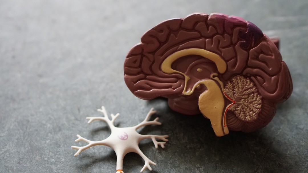
One of the most interesting qualities of gliomas is that they rarely spread to areas outside of the brain and spinal cord. This finding suggests that there is something unique about the microenvironment of the brain and spinal cord that supports the growth of these tumours. Collectively, the brain and spinal cord are known as the central nervous system (CNS).
CNS Microenvironment
Microenvironment refers to the small-scale environment that surrounds cells in different tissues of the body. The microenvironments of different tissues have similarities and differences that uniquely impact the functioning of cells in a particular tissue.
The brain microenvironment is composed of many cell types. Three of the main cell types are: neurons, astrocytes and oligodendrocytes. Neurons are the main messenger cell type of the brain. They facilitate the communication between the body and brain (and vice versa), to enable movement, thought, sensation and general functions necessary for life. Astrocytes and oligodendrocytes are supporting cells that aid in the functioning of neurons.

One of the interesting things about neurons is that they communicate with each other and with target tissues in the body through both electrical and chemical signals. Chemical signals (known as neurotransmitters) are used to communicate between neurons. Electrical signals (known as action potentials) are used to pass the signal through a single neuron. Together, these signals allow neuron-neuron communication through the following steps:
- Neurotransmitter is released from the axon terminus region of a neuron.
- Neurotransmitter travels through the space between 2 neurons to reach the target neuron.
- Neurotransmitter binds to a receptor on the neuronal branching structures known as dendrites (where chemical signals are always received), initiating an action potential (AP – an electric impulse that sends a signal).
- AP travels along the neuronal axon until it reaches the axon terminals.
- At the axon terminals, the AP initiates neurotransmitter release and the cycle repeats.

This unique method of communication between neurons impacts the brain microenvironment. In addition to being comprised of various cell types, neurotransmitters and other by-products of this communication are also important components. Recently, the research community has taken more of an interest in understanding how the brain microenvironment affects cell function, especially that of brain tumour growth.
In 2015, researchers showed that electrically activated neurons released a protein called neuroligin-3, which was found to stimulate the growth of high-grade glioma cells. This result demonstrated that normal brain electrical activity had the potential to impact cancer growth.
As a follow up to this work, two separate research groups delved deeper into the idea that the normal brain surrounding a tumour actually helps gliomas to grow. In 2019, both groups published results demonstrating that there is a close functional relationship between glioma cells and normal neurons. It was found that cancer cells from gliomas (but not other brain tumours) were capable of forming functional connections with surrounding normal neurons. Interestingly, these connections were unidirectional, always passing information from neuron to glioma and never in the opposite direction.
To make things even more interesting, once the signal was transmitted from neuron to glioma, the glioma cells appeared to spread the electrical stimulus amongst each other. As it turns out, the glioma cells were forming physical connections with each other. This physical connection allowed a large network of glioma cells to share the signals they received from neurons.
But what do these signals do once they reach glioma cells?
Using several experimental methods, it was demonstrated that the electrical signals received from neurons stimulated the growth of glioma cells. What’s more, blocking the area that is used by glioma cells to receive the stimulus from neurons stopped glioma growth. Together, these experiments cemented the pro-growth effect that neuronal activity has on glioma cells. This discovery was important because it revealed a potential new mechanism that could be used therapeutically to inhibit glioma growth – by blocking neuronal-glioma communication!
In a final experiment, Dr. Humsa Vanketesh and their team aimed to assess whether neuronal-tumour electrical communication could be detected in human subjects with high-grade gliomas. To perform this experiment, electrodes were placed on the different areas of the brain during surgery to measure neuronal activity. The electrodes were placed at the tumour core, the tumour-normal brain border and the normal brain. They found that the level of neuronal activity was highest at the tumour-normal brain border, suggesting that this functional electrical relationship also exists in intact human tumours.
Together, these results identified a new interaction between the normal brain microenvironment and glioma cells. Although these findings did not directly identify a new therapy, they did deepen our understanding of how glioma cells survive and thrive in the brain.
Hopefully, as we discover more of the intricate processes that keep glioma cells alive, we can identify more weaknesses that can be used to treat this devastating disease.
References
- Claus, EB et al. (2015) Survival and low-grade glioma: the emergence of genetic information. Neurosurg Focus
- Bahadur, S et al. (2019) Current promosing treatment strategy for glioblastoma multiforme: a review. Oncol Rev
- Thomas, A and Noel, G. (2019) Medulloblastoma: optimizing care with a multidisciplinary approach. J Multidiscip Healthc
- Claes, A, Idema, AJ and Wesseling, P. (2007) Diffuse glioma growth: a guerilla war. Acta Neuropathol
- Vankatesh, HS et al. (2017) Targeting neuronal activity-regulated neuroligin-3 dependency in high-grade glioma. Nature
- Venkataramani, V et al. (2019) Glutamatergic synaptic input to glioma cells drives brain tumour progression. Nature
- Vankatesh, HS et al. (2019) Electrical and synaptic integration of glioma into neural circuits. Nature
